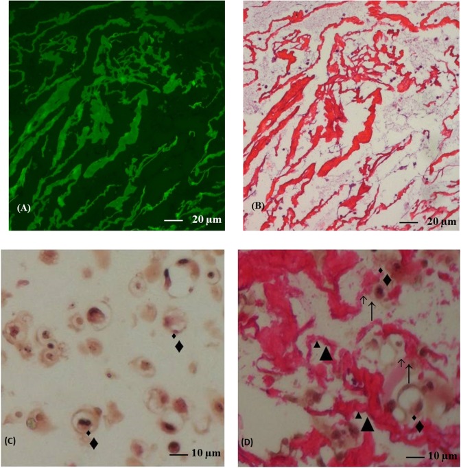
Fig 3. Hematoxylin and eosin stained section from alginate and chitosan-alginate beads.
Fluorescence microscopy in chitosan-alginate beads section (A). The same section stained by hematoxylin and eosin (B; original magnification x20). Section in alginate (C) or chitosan-alginate (D) beads stained by hematoxylin and eosin (original magnification x40). Black triangles represented chitosan trabeculae, black diamonds represented chondrocytes and ↑showed contact.

