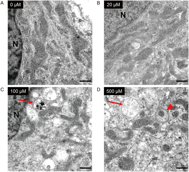
Fig 10. Effect of ketamine on mitochondrial morphology in neurons derived from iPSCs.
Cells were treated with ketamine for 24 h, and observed by transmission electron microscopy. Untreated cells (A) and cells treated with 20 μM ketamine (B) had elongated mitochondria with intact inner and outer membranes. Treatment with 100 μM ketamine (C) resulted in fragmented mitochondria and the presence of autophagosomes (arrow). After treatment with 500 μM ketamine (D), the structure of the mitochondria became discrete and round, and the mitochondrial length was shortened. Autophagosomes (arrow) were detected, and some fragmented mitochondria were degraded by autophagosomes (arrowhead). N: nucleus. Scale bar = 500 nm.

