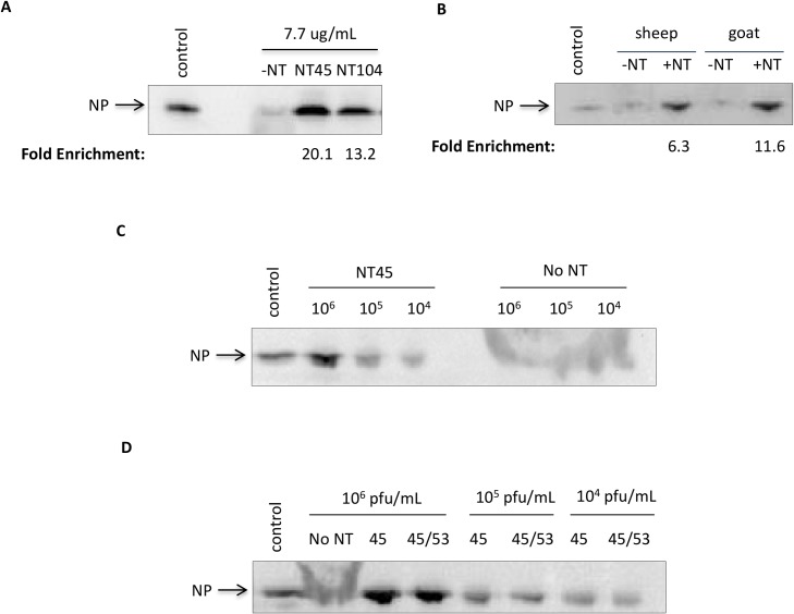
Fig 6. Nanotrap particles can capture and enrich NP found in various animal sera.
A) Cytoplasmic extracts from RVFV infected cells were diluted to 7.7 μg/ml in 100% sheep serum. One milliliter of sample was incubated with 100 μl of NT45 or NT104 for 30 minutes. No Nanotrap particle (-NT), control (CE at 77.7 μg/ml diluted in 50mM Tris-HCl), and mock-infected samples at 10 μl volumes were processed in parallel. The (+)NT samples were then centrifuged and washed once with 0.25M sodium thiocyanate and twice with diH2O. The samples were analyzed by western blotting for NP using antibodies against NP. B) Cytoplasmic extracts from RVFV infected cells were diluted to 7.7 μg/ml in 100% sheep, goat, or donkey sera. One milliliter of sample was incubated with 100 μl of NT45 for 30 minutes. No Nanotrap particle (-NT) and control (CE at 7.7 μg/ml diluted in 50mM Tris-HCl) samples at 10 μl volumes were processed in parallel. The samples were processed as in panel A. C) Viral supernatants were diluted in sheep serum for final viral titers of 1E+06 pfu/ml, 1E+05 pfu/ml, and 1E+04 pfu/ml. One milliliter of the sample was incubated with 100 μl of NT45 for 30 minutes. Control sample is viral supernatant at 1E+07 pfu/ml (10 μl volume). The samples were processed as in panel A. D) Viral supernatants were diluted in sheep serum for final viral titers of 1E+06 pfu/ml, 1E+05 pfu/ml, and 1E+04 pfu/ml. One milliliter of the sample was incubated with 100 μl of NT45 or 100 μl of equal parts NT45 and NT53 for 30 minutes. Control sample is viral supernatant at 1E+07 pfu/ml (10 μl volume). The samples were processed as described in panel A.

