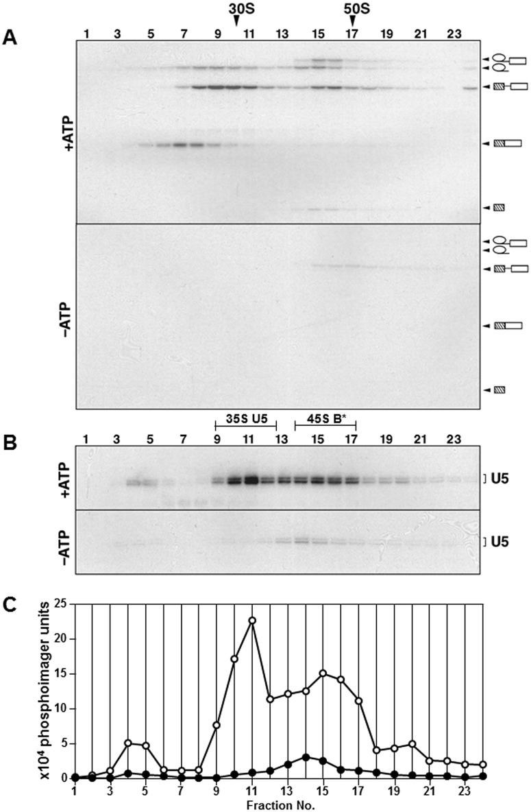
Fig 3. Characterisation of the complexes released into the supernatant from immobilised activated spliceosomes during splicing in the presence or absence of ATP by glycerol gradient centrifugation.
(A) The RNA extracted from each gradient fraction was fractionated by denaturing PAGE and the 32P-containing species were detected by autoradiography. S-values were determined by comparison with the reference gradients containing 30S and 50S ribosomal subunits. The RNA identities are shown on the right. (B) Northern blot analysis of gradient fractions from panel A with the U5 snRNA specific probe. Fractions corresponding to the 35S U5 snRNP and 45S B* spliceosomes are underlined. (C) Quantification of the signals corresponding to the U5 snRNA (panel B) using a PhosphorImager. Open circles correspond to the reaction carried out in the presence of ATP and closed circles—in the absence of ATP.

