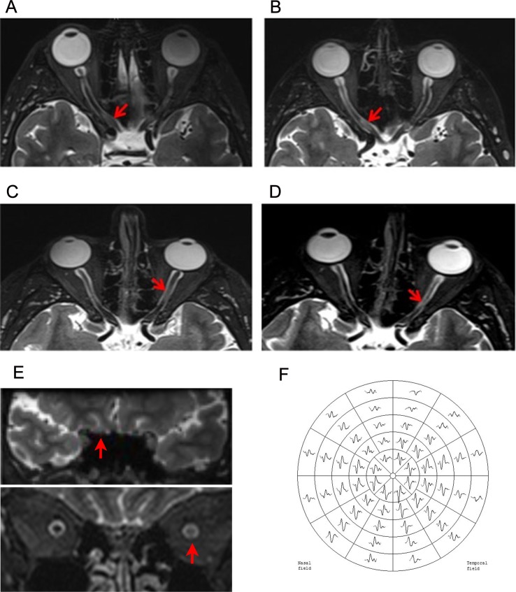
Fig 1. Example of optic nerve MRI lesions after ON and mfVEP recording.
A-B: Intra-cannalicular lesion at 1 month (A) and 12 months (B) after ON. Lesion length decreased from 9.9 mm to 7 mm. C-D: Intra-orbital lesion at 1 month (C) and 12 months (D) after ON. Lesion length decreased from 11.3 mm to 5.5 mm. E: Coronal views of intra-cannalicular (above) and intra-orbital (below) lesions. F: Example of mfVEP trace.

