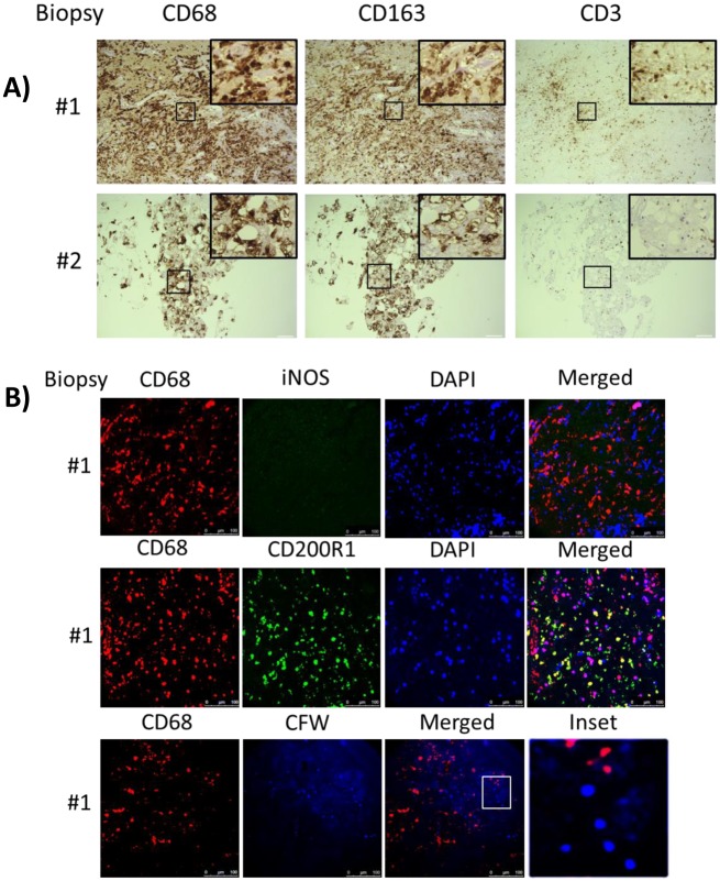
Fig 6. Brain biopsy specimens from patients with s-CNS cryptococcosis demonstrate macrophage and T-cell tissue infiltration.
A) Diagnostic specimens obtained from two s-CNS patients were stained with macrophage markers CD 68 and CD163 and T-cell marker CD3. B) Immunofluorescence of brain biopsy of one patient after staining with macrophage marker CD68 and M1 marker (iNOS) and M2 marker (CD200R1), fungal stain, calcofluor white (CFW) and nuclear stain, 4',6-diamidino-2-phenylindole (DAPI). Magnification at 10 x with inset at 20x and scale bar set at 100 μm for 10x images.

