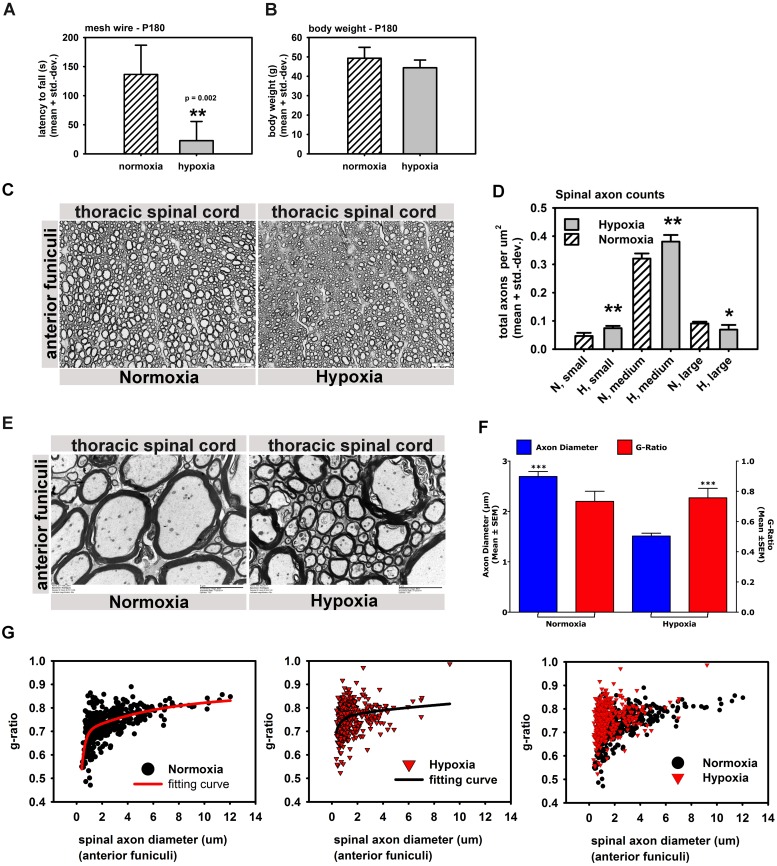
Fig 5. Abbreviated hypoxia changes axonal composition in spinal cords and causes dysmyelination of spinal axons.
A, B: 6 month old hypoxic mice showed strong motor deficits in hanging wire tests (A) without having different body weights (B) (n = 4 normoxic + 6 hypoxic animals). C: Spinal cord thick sections from anterior and lateral funiculi (thoracic regions) in hypoxic and normoxic mice 6 month after the hypoxic insult (4 control + 4 hypoxic mice). D: Automated axon counts of spinal cord thick sections from C. showing abnormal axonal compositions in spinal anterior funiculi with increased small and medium diameter axons and decreased numbers of large diameter axons relative to normoxic controls (small = 1–4 μm; medium = 4–10 μm; large = > 10 μm) (4 control + 4 hypoxic mice). E: Electron microscopy of spinal cord sections left and right of the anterior median fissure (thoracic spinal cord) in hypoxic and control animals (magnification: 8kx). Higher g-ratios (thinner myelin sheaths) and loosely wrapped myelin around axons was prominent by Electron microscopy in thoracic spinal motor neurons (anterior funiculi) (4 control + 4 hypoxic mice, 100 axons per animal). F. Chi-Square analysis of the g-ratio/axon diameter relationship in hypoxic and normoxic animals. G: Scatter plots of g-ratio vs. axon diameter in control (black) and hypoxic animals (red) with best fitting curves, with *** equals p < 0.001; ** equals p < 0.01; * equals p < 0.05.

