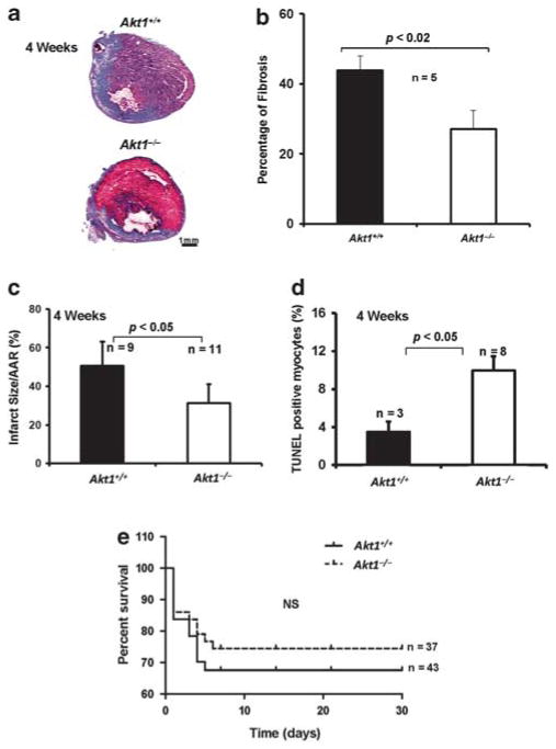
Figure 4.
Improved cardiac remodeling and enhanced survival rate in Akt1−/− mice 4 weeks after MI. (a) Representative images of the Akt1+/+ and Akt1−/− mice heart sections stained with Masson’s trichrome staining showing fibrosis 4 weeks after MI. (b) Quantification of the fibrotic area in mice hearts represented as mean percentage of fibrosis±s.e.m. indicating significantly larger fibrotic area 4 weeks after MI in Akt1+/+ hearts compared with Akt1−/− hearts (n =5/group). (c) Quantification of the infarct area in Akt1+/+ and Akt1−/− mice hearts 4 weeks after MI represented as mean±s.e.m., indicating significantly lesser infarct area in Akt1−/− mice compared with Akt1+/+ mice (n =9–11). (d) Quantification of the TUNEL-positive cells in Akt1+/+ and Akt1−/− mice hearts 4 weeks after MI (n =3–8) represented as mean apoptotic area±s.e.m. indicating significantly higher number of apoptotic cells in Akt1−/− hearts compared with Akt1+/+. (e) Akt1+/+ and Akt1−/− mice were subjected to MI or sham surgery and survival was monitored for 30 days and analyzed by the Kaplan–Meier method (n =37 and 43).

