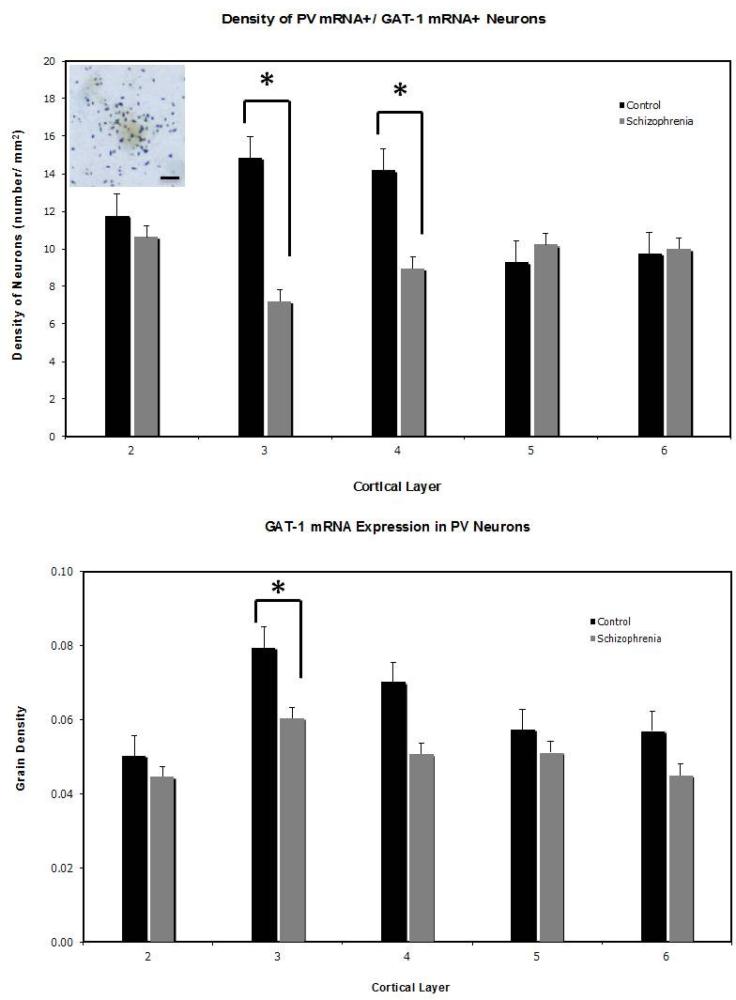
Figure 1.
Upper panel: Mean (±SEM) density of PV+/GAT-1+ neurons is significantly decreased in layers 3 and 4 in the PFC in schizophrenia. Photomicrograph shows a double-labeled (PV+/GAT-1+) neurons. Scale bar = 10 μm. Lower panel: Mean (±SEM) density of silver grains over PV+ neurons is significantly decreased in layer 3 in the PFC in schizophrenia. Layer 1 was not included in the analyses because no PV+ neurons were found in this layer. *p<0.05, based on post-hoc group comparisons.

