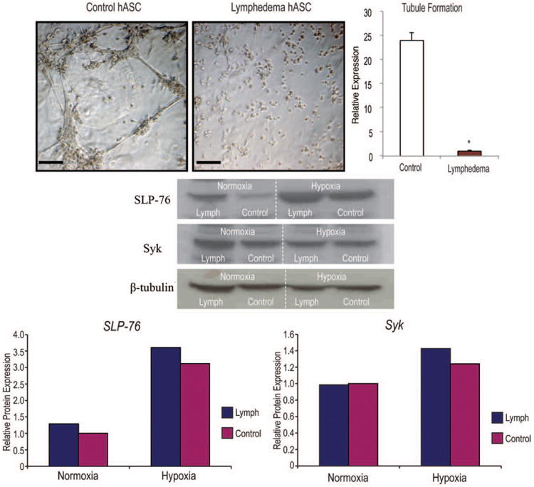
Fig. 4.

Lymphedema-associated adipose-derived stem cells are less vasculogenic than healthy control adipose-derived stem cells. (Above, left) In vitro Matrigel tubule formation assay of lymphedema-associated and control adipose-derived stem cells after 12 hours of hypoxia (1% oxygen). (Above, right) Tubule formation was quantified and counted by three blinded, independent observers. (Center) Western blot analysis of lymphedema-associated and control adipose-derived stem cells cultured either under normoxic or hypoxic (1% oxygen) conditions for 24 hours (Below). SLP-76 and Syk were analyzed and normalized to β-tubulin. Two-tailed t test was used to compare groups (mean expression ± SD, *p < 0.05). hASC, human adipose-derived stromal cells. Quantification was performed using densitometry and ImageJ (National Institutes of Health, Bethesda, Md.).
