Figure 2.

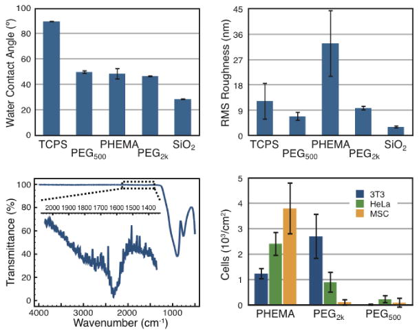
(a) Water contact angle (n=5) determined with PBS (pH=7.4). (b) Root mean squared (RMS) surface roughness (n=3) determined using atomic force microscopy (AFM). (c) FTIR characterization of PEG500-coated substrates demonstrating carbonyl stretching from the carbamate functionality arising from the isocyanate coupling and the methacrylate functionality. (d) Cell adhesion assays with NIH 3T3 fibroblast, HeLa, and mesenchymal stem (MSCs) cells demonstrating negligible adhesion of cells to PEG550-coated substrates (3T3 = (0.01 ± 0.02) ×103 cells/cm2, HeLA = (0.22 ± 0.2) ×103 cells/cm2, MSCs = (0.08 ± 0.2) ×103 cells/cm2).
