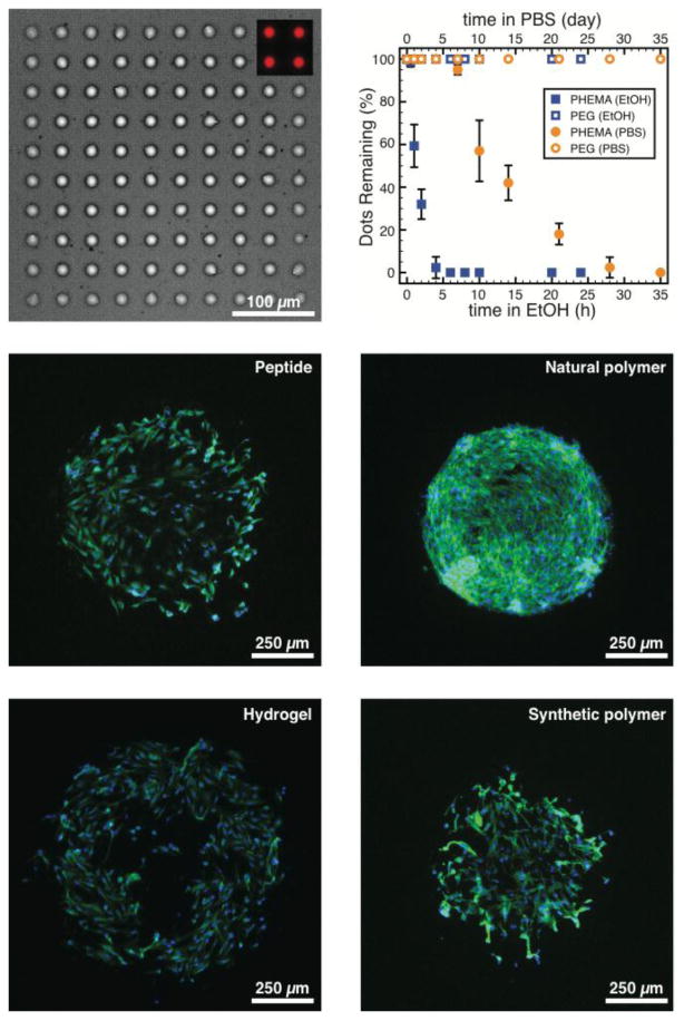
Figure 3.

(a) Microscopy images (bright-field) of large (∼ 25μm) polymer dots composed of PEG acrylate (PEGA; 30%; Mn = 480 Da) and trimethylolpropane triacrylate (TMPTA; 70%) printed in 10 × 10 arrays in triplicate on PEG500-coated substrates. Polymer dots contain rhodamine-functional acrylate comonomer imparting strong red fluorescence (inset). (b) Stability of the printed polymer dots was investigated over time in ethanol (20°C, lower axis) and PBS (pH=7.4, 37°C, upper axis). Representative fluorescence microscopy images of 3T3 fibroblast cells grown on functional spots (d ∼ 500 μm; n=20) composed of (c) RGDC peptides (printed at 100 μg/mL), (d) poly(d-lysine) (printed at 100 μg/mL), (e) polyacrylamide hydrogel containing collagen I (0.05%), and (f) PEGA-co-TMPTA (as in part a and b), printed on PEG500-coated substrates.
