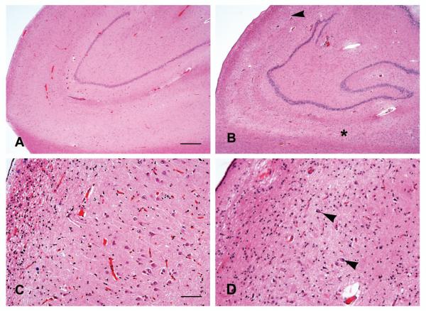
Figure 1.
Photomicrographs showing the CA1 and subiculum in cases without (A) and with (B) HS in which sclerosis involves CA1 (starting below the arrowhead) and the subiculum (right of the *) in continuity. Higher magnification shows preservation of pyramidal neurons in the CA1 in the absence of sclerosis (C) and astrogliosis with few residual neurons (arrowheads) in the case with HS (D). Hematoxylin-eosin stain, Scale bar = 500 μm (A,B) and 200 μm (C, D).

