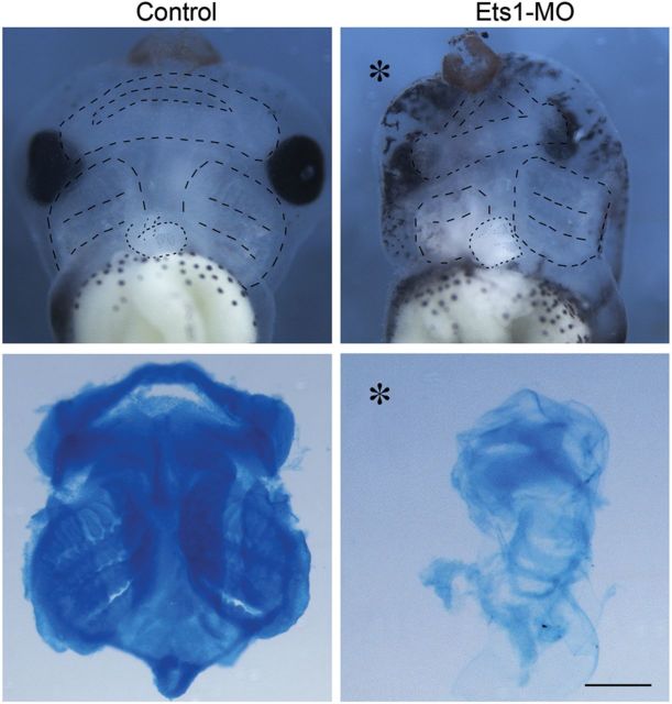
Figure 6.
Ets1-MO disrupts cranial cartilage formation. Ventral views of the stage 45 heads illustrate formation of cranial cartilage outlined in upper panels. Alcian blue stained cartilage were dissected and shown in the lower panels. While mandibular, hyoid, and posterior branchial arch cartilages were distinct in the control head (n = 12), cartilage formation was completely abolished by Ets1-MO (asterisk marks the injected half; n = 14). Cartilage formation in the contralateral side of Ets1-MO injected embryo was also impaired, likely due to a delay in cartilage maturation. Scale bar = 0.5 mm.

