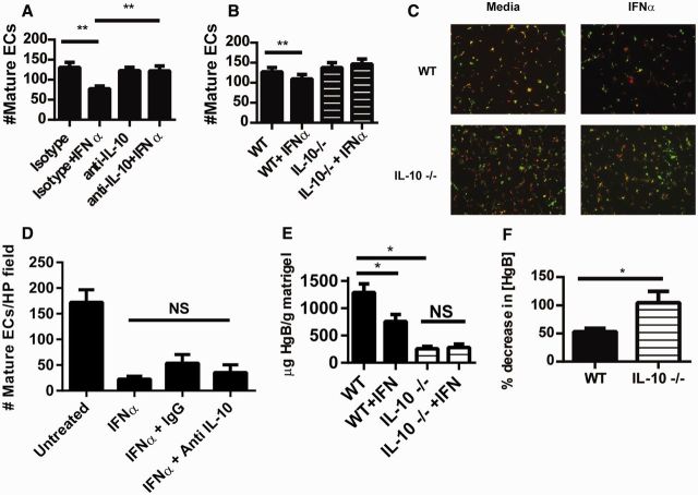
Fig. 2.
IL-10 is required for the inhibitory effects of type I IFN on murine EPC differentiation and in vivo neo-angiogenesis
(A) Wild-type (WT) murine EPCs were incubated with 1000 U/ml IFN-α in the presence or absence of a neutralizing IL-10 antibody or isotype control. Following 7 days of culture, mature ECs were quantified as in Fig. 1 using BS-1 instead of UEA-lectin. Results represent the mean (s.e.m.) of mature ECs quantified in triplicate from three different mice. (B) WT (n = 4) or IL-10−/− (n = 6) EPCs were incubated with IFN-α and mature ECs were quantified as in Fig. 2A. (C) Representative images (total magnification 100 × ) of murine EC cultures from Fig. 2B (green: BS-lectin; red: ac-LDL). (D) Human EPC/CAC cultures (n = 6) were treated as in Fig. 2A utilizing a neutralizing antibody specific for human IL-10. On day 14 of culture, mature ECs were quantified as in Fig. 1. **P ≤ 0.01, NS: non-significant. (E and F) Matrigel embedded with 20 nM fibroblast growth factor with or without 1000 U/ml IFN-α was injected subcutaneously into WT (n = 13) or IL-10−/− (n = 8) mice. Seven days after injection, Matrigel plugs were harvested and their HgB content was quantified as an estimate of neo-angiogenesis within the plug. Results in Fig. 2A represent HgB quantification. Results in Fig. 2B show the percentage inhibition of angiogenesis in plugs containing IFN-α as compared with plugs not exposed to IFN-α. *P < 0.05. BS-1: Bandeiraea (Griffonia) simplicifolia lectin 1; CAC: circulating angiogenic cell; EPC: endothelial progenitor cell; HgB: haemoglobin; UEA: Ulex europaeus agglutinin 1.

