Figure 1.
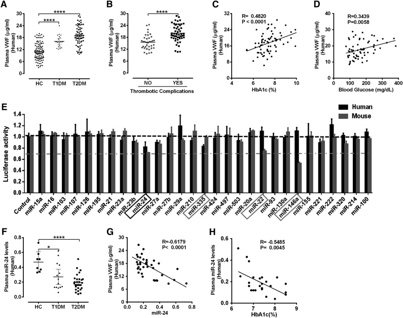
Elevated VWF and reduced miR-24 identified in diabetic plasma. (A) ELISA analysis and comparison of VWF levels in plasma of HCs (n = 112), T1DM (n = 33), and T2DM (n = 82); ****P < .0001. (B) Comparison of mature VWF levels in plasma of T2DM with or without thrombotic complications (acute coronary syndrome and cerebrovascular events). ****P < .0001. (C) Correlation between plasma VWF levels and HbA1c (%) levels. (D) Correlation between plasma VWF levels and blood glucose level. (E) A panel of 29 candidate VWF-targeting miRNAs (including 25 highly expressed miRNAs in endothelial cells and 4 miRNAs predicted by TargetScan) cotransfected with VWF 3′UTR Lenti-reporter-Luc vector into 293 T cells. The luciferase activity was measured and normalized to empty control reporter and empty miRNA control. miR-24, miR-335, miR-22 and miR-146a (highlighted) demonstrated a significant reduction in luciferase activity. These assays were performed in quadruplicate and repeated 3 times. (F) qPCR analysis of circulating miR-24 levels (normalized to cel-miR-238 spike-in) in plasma of healthy controls (n = 8), T1DM (n = 14), and T2DM (n = 30); ****P < .0001; *P < .05. (G) Correlation between plasma VWF levels and circulating miR-24 in DM (n = 37). (H) Correlation between circulating miR-24 and HbA1c (%) in DM (n = 25).

