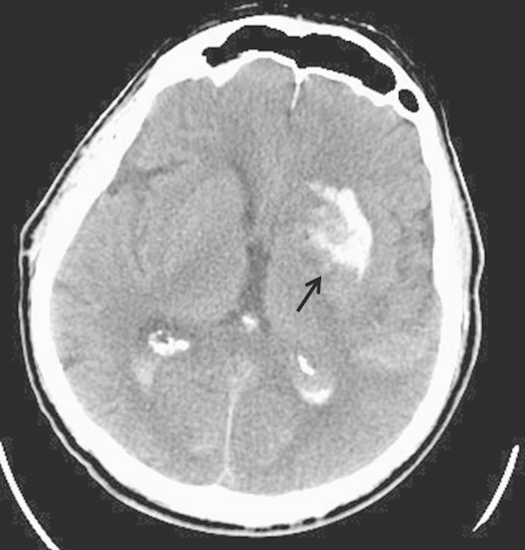
Fig. 2.

Axial noncontrast CT scan following thrombectomy for internal carotid artery occlusion and ischemic stroke. A “contrastoma” has appeared in the left thalamus due to extravasation of contrast material secondary to blood–brain barrier breakdown (arrow).
