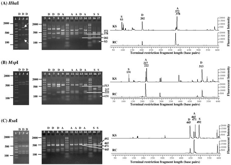
FIG. 2.
Restriction digest analysis of selected cloned 16S rRNA gene fragments with HhaI (A), MspI (B), and RsaI (C) and detection of community members in T-RFLP profiles. The letters D, A, and S indicate Dehalococcoides, Acetobacterium, and Desulfuromonas clones, respectively. Lanes 1, 5, 10, and 15, 50- to 2,000-bp ladder; lane 2, Dehalococcoides sp. strain FL2; lane 3, Dehalococcoides ethenogenes strain 195; lane 4, Dehalococcoides sp. strain CBDB1; lane 6, ARC13 (D); lane 7, ARC18 (D); lane 8, ARC61 (D); lane 9, ARC62 (A); lane 11, AKS04 (A); lane 12, AKS17 (A); lane 13, AKS31 (D); lane 14, AKS48 (A); lane 16, AKS67 (S); lane 17, AKS68 (S). The white arrows (A) point to the unique band obtained for Dehalococcoides ethenogenes strain 195 when the cloned 16S rRNA gene was digested with enzyme HhaI as well as the missing diagnostic band at 202 bp. The numbers to the right of the gel images indicate the sizes of the corresponding fragments also detected in the T-RFLP profiles. The numbers indicated are the actual T-RF lengths and not the longer restriction fragments obtained due to an additional portion (27 or 55 bp) of the TA cloning vector.

