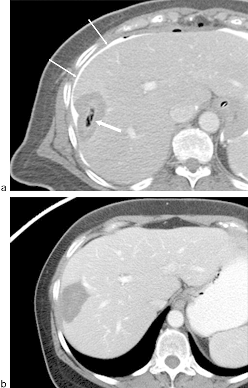
Fig. 1.

Gas in the region of ablation following radiofrequency ablation (RFA) of a liver metastasis from breast carcinoma. (a) Axial image from a CT scan with contrast immediately following RFA demonstrates gas within the zone of ablation (arrow). Because of the location of the metastasis adjacent to the liver capsule and chest wall, a solution of 5% dextrose water and iodinated contrast was instilled in the perihepatic space (thin arrows). Note that the gas bubbles do not correspond to the ablation margins. (b) Axial CT image with contrast 3 months following ablation demonstrates resolution of gas. There is also no evidence of enhancing tissue to suggest residual disease.
