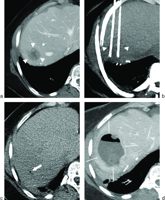
Fig. 2.

Hepatic abscess following radiofrequency ablation (RFA) of liver metastasis from pancreatic neuroendocrine carcinoma. The patient had previously undergone a Whipple procedure 2 years prior to RFA. (a) Axial image from a CT scan with contrast demonstrates a hypodense lesion near the hepatic dome (arrowheads) consistent with a hepatic metastasis. (b) Thick-slab reformatted axial CT image during ablation demonstrates the RFA probes along the medial and lateral aspects of the lesion (arrowheads). (c) Axial CT image without contrast immediately following ablation demonstrates a small focus of gas (arrow), which is normal following liver RFA. (d) Axial CT image with contrast performed 3 weeks following RFA demonstrates findings of a hepatic abscess following RFA (thin arrows): rim enhancement, low-density center, and increasing intralesional gas.
