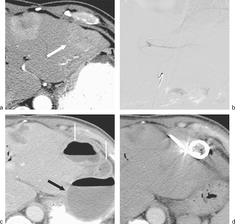
Fig. 4.

Hepatic abscess following transarterial bland embolization for metastatic neuroendocrine cancer. The patient had previously undergone a Whipple procedure 3 years prior to embolization. (a) Axial image from a CT scan during the arterial phase on enhancement demonstrates a hyperenhancing lesion in the left hepatic lobe (arrow) consistent with a hepatic metastasis. (b) Bland particle embolization of the segment 2 and 3 left hepatic artery was performed with 355–500 µm polyvinyl alcohol particles. Image from a digital subtraction angiogram demonstrates no significant residual enhancement of the left hepatic lobe lesion. (c) Axial CT image with contrast performed 3 weeks following RFA demonstrates findings of a complex hepatic abscess (thin arrows). A large extrahepatic abscess was also present (black arrow). The complex intrahepatic and extrahepatic abscesses were drained percutaneously. (d) Axial CT image performed 2 weeks following percutaneous drainage demonstrates interval resolution of both abscesses.
