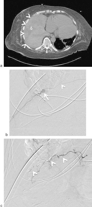
Fig. 4.

A 67-year-old woman developed severe hypotension and tachycardia following surgical chest tube placement for empyema at an outside hospital. Chest tube was noted to be draining bright red bloody fluid. (a) Axial image from an emergent CT scan revealed a large right hemothorax (arrowheads). (b) Selective catheterization (arrowhead = microcatheter) of the right ninth intercostal artery. Angiography revealed a pseudoaneurysm of the intercostal artery (arrow). (c) Successful coil (arrowheads) embolization of the intercostal artery distal and proximal to pseudoaneurysm.
