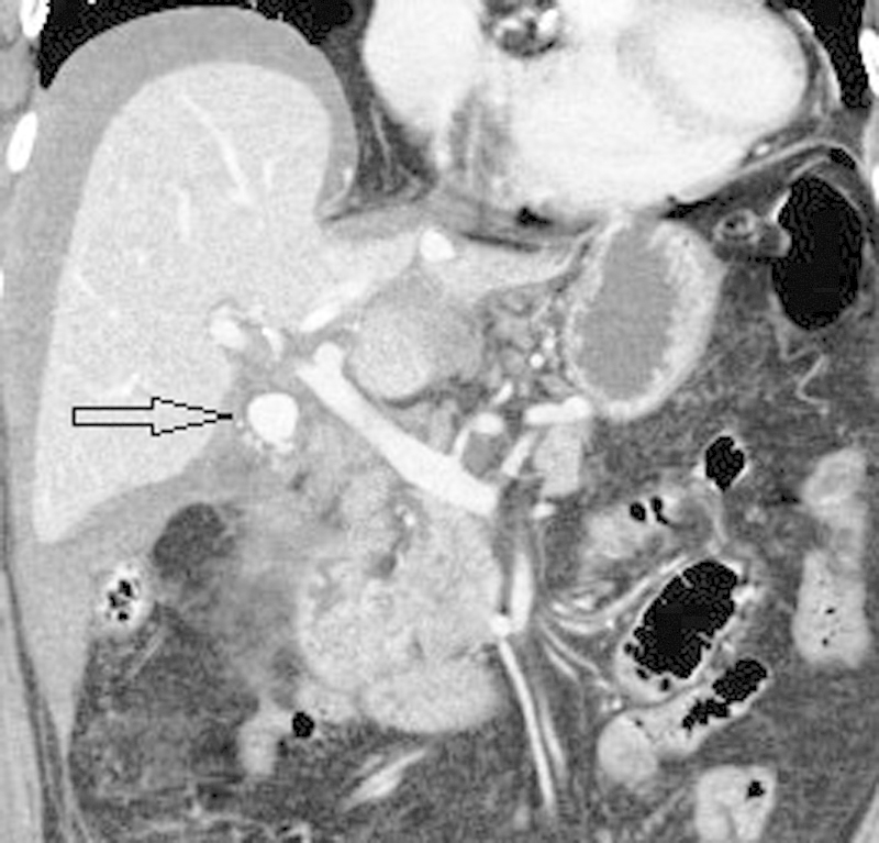
Fig. 10.

An 82-year-old woman presented to the emergency room with sudden onset of abdominal pain, tachycardia, and hypotension 3 days after laparoscopic cholecystectomy. Contrast-enhanced coronal CT scan image shows a loculated collection of contrast measuring ∼1.7 × 1.5 cm in the gallbladder fossa (arrow) representing a pseudoaneurysm. There is high-density perihepatic fluid due to intra-abdominal hemorrhage. The pseudoaneurysm was successfully treated by coil embolization of the cystic artery.
