
Fig. 4.
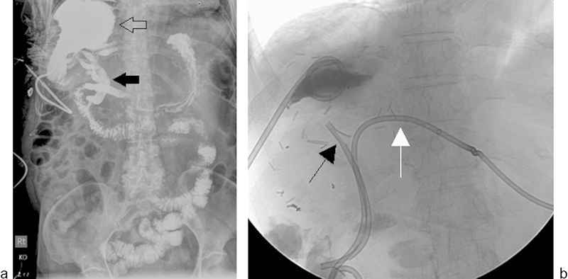
A 72-year-old woman underwent right hepatectomy and resection of the caudate lobe for hepatocellular carcinoma. On postoperative day 2, a large fluid collection was identified in the surgical bed. The collection was drained by a 12 French drainage catheter placed under CT guidance (not shown). Large volume drainage from the catheter persisted for ∼2 months. (a) Fluoroscopic injection of the large right subphrenic fluid collection cavity (open arrow) shows fistulous communication with the biliary system (solid arrow). (b) Additional drainage by placement of an endoscopic biliary stent (black arrow) and left internal–external biliary drain (white arrow) resulted in complete resolution of communication after 6 weeks. Separate contrast injection of the right upper quadrant abscess drainage catheter and biliary catheter revealed no residual communication between the right perihepatic fluid collection and the bile ducts (not shown).