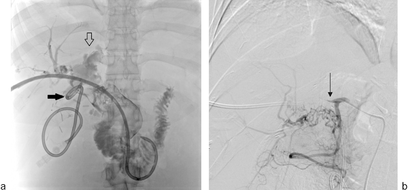
Fig. 7.

A 38-year-old woman with carcinoid tumor metastases to the liver underwent extended left hepatectomy, hepatic artery thrombectomy with interposition graft, and choledochocholedochostomy. The patient had persistent bile leak from the T-tube tract and underwent PTHC. (a) PTHC shows complete loss of integrity of the common bile duct (open arrow), which is replaced by debris. A pigtail catheter is seen in the liver draining a biloma (solid arrow). (b) Digital subtraction angiography of the proper hepatic artery performed via a right common femoral artery approach demonstrates complete occlusion of the hepatic artery (arrow).
