Figure 5.
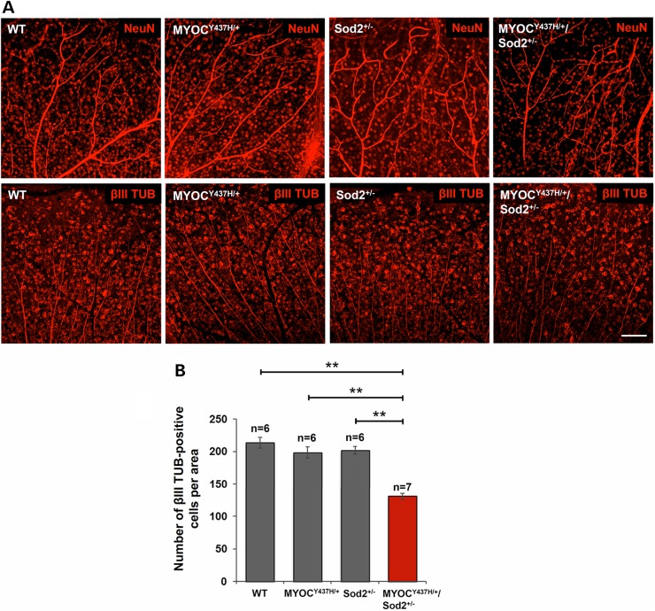
Loss of RGCs in the peripheral retinas of Tg-MYOCY437H/Sod2+/− mice. (A) RGCs were labeled by anti-NeuN (upper row) and anti-βIII TUB (lower row) antibodies in the whole-mounted retinas of 10-month-old littermates. Representative images from the peripheral retinas are shown. Scale bar, 100 μm. (B) βIII TUB-positive cells in the peripheral retinas of 10- to 12-month-old mice were counted and plotted as the numbers per area. All the values are presented as mean ± SE and analyzed by one-way ANOVA followed by Tukey's multiple comparisons test. **P < 0.01; n shows the number of analyzed mice.

