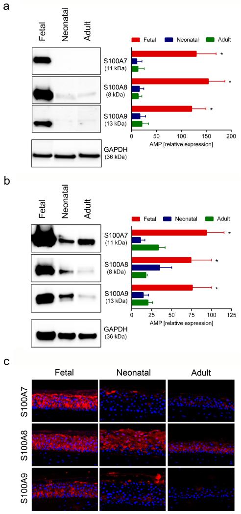
Figure 3. Fetal KC express high levels of S100A7, S100A8 and S100A9 protein in monolayer and SE cultures.
Fetal, neonatal and adult KC were cultured under proliferating conditions in monolayer cultures and analysed for the expression of S100 proteins (a). Western blot analysis of KC showed higher expression of S100 proteins in fetal KC, as compared to neonatal and adult KC. Western blots of representative samples and densitometric analysis of four to six independent experiments in each group are shown. *p<0.05 for fetal KC compared to neonatal and adult KC; SE from fetal, neonatal and adult KC were established and analyzed by Western blot analysis (b) and immunfluorescence staining (c). Western blot analysis showed higher expression of S100 proteins in SE from fetal KC, as compared to neonatal and adult KC. Western blots of representative samples and densitometric analysis of five to seven independent experiments in each group are shown. *p<0.05 for fetal KC compared to neonatal and adult KC. (c) Immunofluorescence staining of SE cultures showed higher expression of S100 proteins in SE cultured from fetal KC as compared to neonatal and adult KC; one representative experiment out of three is shown.

