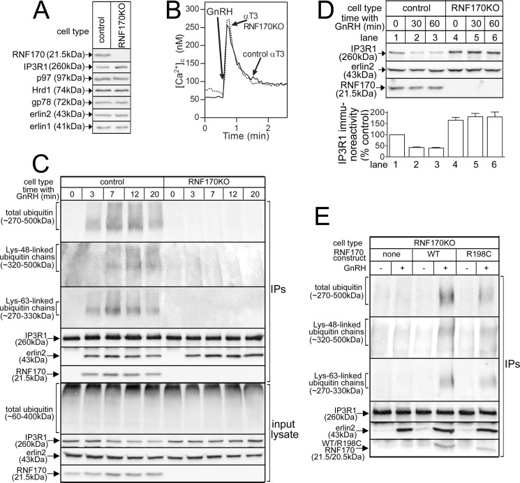
FIGURE 7.
CRISPR/Cas9-mediated deletion of RNF170 and reconstitution with exogenous RNF170 constructs. A, levels of RNF170, IP3R1, and other pertinent proteins in lysates from αT3 control and RNF170KO cells. B, GnRH (0.1 μm)-induced Ca2+ mobilization in αT3 control and RNF170KO cells. C, IP3R1 ubiquitination in αT3 control and RNF170KO cells. Cells were incubated with 0.1 μm GnRH and anti-IP3R1 IPs and input lysates were probed for the proteins indicated. D, IP3R1 down-regulation in αT3 control and RNF170KO cells. Cells were incubated with 0.1 μm GnRH and lysates were probed for the proteins indicated. The histogram shows combined quantitated immunoreactivity (mean ± S.E., n = 4). E, reconstitution of IP3R1 ubiquitination in RNF170KO cells. αT3 RNF170KO cells stably expressing WTRNF170 or R198CRNF170 were incubated without or with 0.1 μm GnRH for 20 min, and anti-IP3R1 IPs were probed for the proteins indicated.

