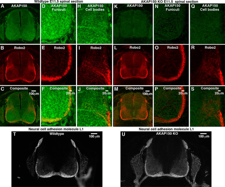
FIGURE 3.
AKAP150 is expressed in the developing spinal cord, where it co-distributes with Robo2 in specialized regions. Transverse sections of embryonic day 11.5 spinal cords from wild type and AKAP150−/− mice were stained for AKAP150 and Robo2 and imaged using wide field fluorescence microscopy (wild type (A–C) and AKAP150−/− (K–M)). AKAP150 is shown in green, and Robo2 is shown in red. Higher magnification spinning disc confocal images provide more detailed localization of these proteins in the funiculi (wild type (D–F) and AKAP150−/− (N–P)) and commissural neuron cell bodies (wild type (H–J) and AKAP150−/− (Q–S)). T and U, sections of spinal cord from wild type (T) and AKAP150−/− mice (U) stained for neural cell adhesion molecule L1, a marker for midline crossing.

