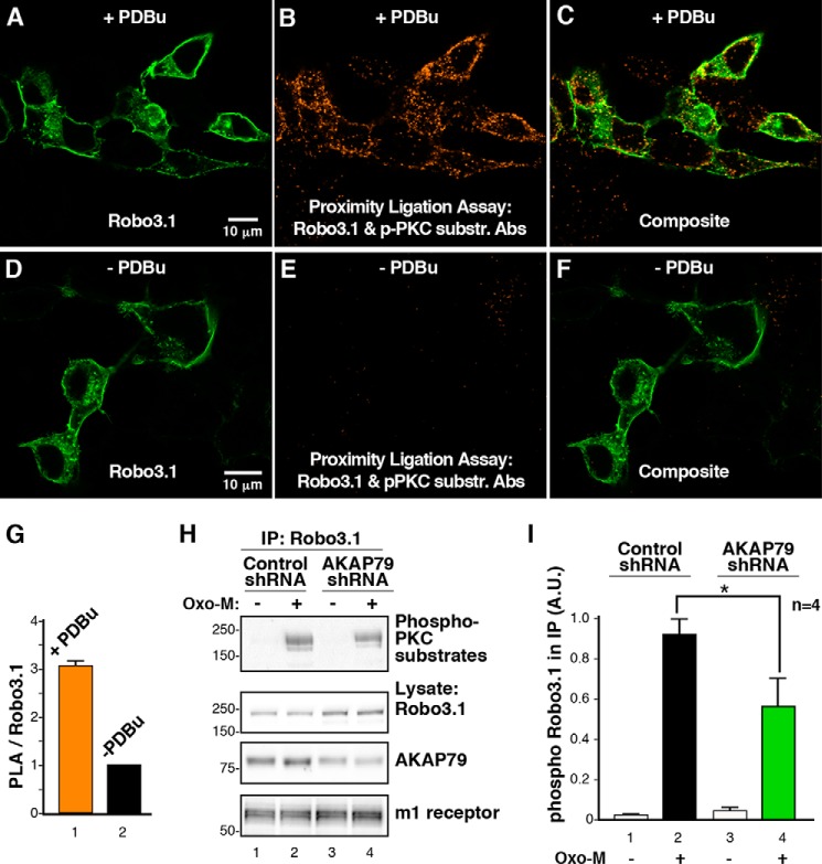
FIGURE 6.
AKAP79 regulates the phosphorylation of Robo3.1 by PKC. A–F, HEK293 cells were transfected with Robo3 and treated with PDBu (A–C) or vehicle (DMSO; D–F) for 10 min. Cells were incubated with a Robo3 antibody and the phospho-PKC substrate antibody, and a PLA reaction was carried out between these two antibodies (B, C, E, and F; orange). Cells were subsequently stained using a V5 antibody to recognize Robo3.1 (A, C, D, and F; green). G, analysis of PLA signal normalized to Robo3 expression (means ± S.E. (error bars), n = 3, >100 cells, p ≤ 0.05, one-sample Student's t test). H, Robo3 and the m1 muscarinic receptor were coexpressed in HEK293 cells with control or AKAP79 shRNA. Cells were treated with vehicle (H2O) or 10 μm Oxo-M for 2 min. V5 immunoprecipitations (IP) of cell lysates were immunoblotted with the PKC substrate antibody (top). Robo3.1 expression (top middle), AKAP79 knockdown (bottom middle), and m1 receptor expression (bottom) were confirmed by immunoblotting. I, quantification of phospho-Robo3 signal present in immunoprecipitations normalized to Robo3 expression (means ± S.E., n = 4, p ≤ 0.05, unpaired Student's t test). A.U., arbitrary units.

