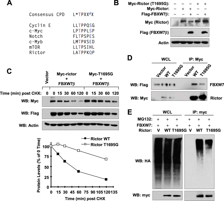
FIGURE 5.
Identification of a putative CPD in rictor (A) and demonstration of its function in FBXW7-mediated rictor degradation (B–E). A, a putative CPD motif was identified at positions 1693–1704 of rictor. B and D, 293T cells were co-transfected with FLAG-FBXW7 and myc-rictor or myc-rictor (T1695G) for 24 h (B) or 48 h (D). The cells were then harvested for preparation of WCLs and subsequent WB (B) or IP/WB (D). C, 293T cells were co-transfected with FLAG-FBXW7 and myc-rictor or myc-rictor (T1695G) and 24 h later treated with 10 μg/ml CHX. At the indicated time points, the cells were harvested for preparation of WCLs. The indicated proteins were detected with WB and quantified with NIH Image J. After being normalized to actin, the results were then plotted as the relative protein levels compared with those at time 0 of CHX treatment (bottom panel). E, FBXW7β was co-expressed with myc-rictor or myc-rictor (T1695G) in 293T cells, and after 24 h, cells were transfected with an empty vector (V) or HA-ubiquitin (HA-Ub). After another 24 h, cells were treated with 10 μm MG132 for 2 h and then subjected to preparation of WCLs and subsequent IP/WB to detect ubiquitinated rictor.

