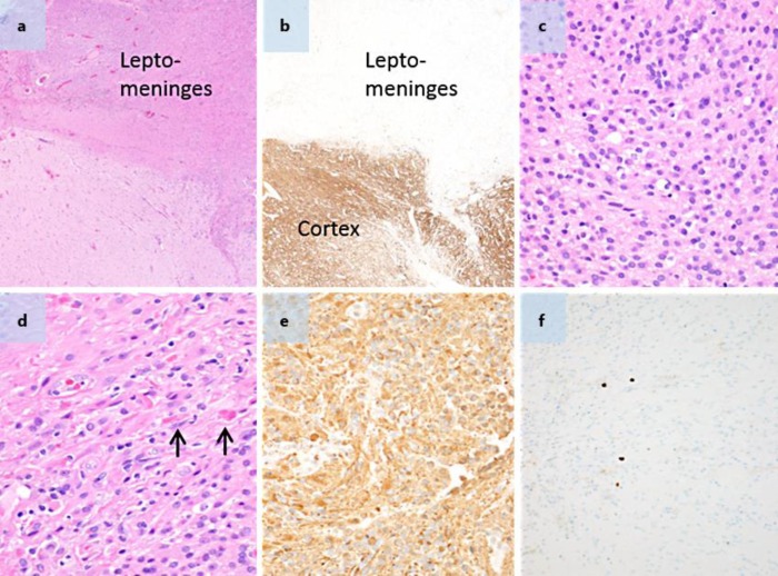
Fig. 3.
a, b HE and neurofilament stains, respectively ×4 objective. The neurofilament stain with paired HE highlights that the tumor predominantly involves the leptomeninges. c HE. ×40 objective. Much of the tumor was composed of monotonous-appearing cells with round and relatively regular nuclei. d HE. ×40 objective. In areas of the tumor, the cells had slightly more elongated cell nuclei, and the tumor contained Rosenthal fibers (arrows). e GFAP. ×40 objective. The tumor cells are strongly positive for GFAP. f Ki-67. ×20 objective. The tumor had a low level of proliferative activity which is highlighted by only occasional scattered Ki-67-positive cells.

