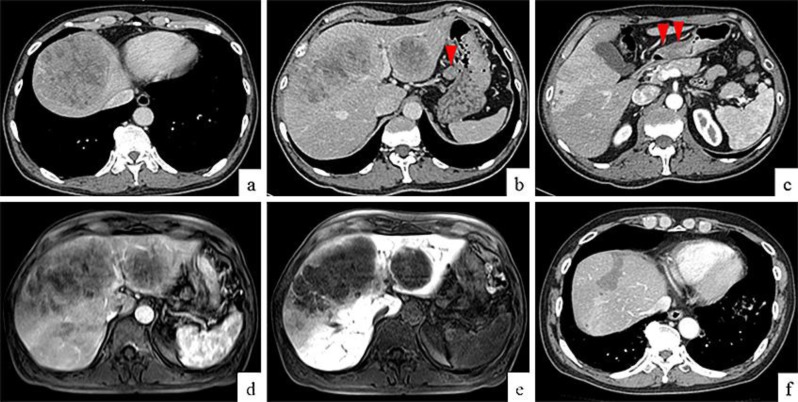
Fig. 1.
CT on admission and 25 months after chemotherapy. Enhanced CT on admission revealed ring enhancement around the periphery of the tumors and heterogeneous enhancement within the tumors (a). CT also revealed multiple hepatic tumors with heterogeneous enhancement, and a swollen lymph node surrounding the lesser curve of the stomach (b, arrowhead). Thickening of the stomach wall was indicative of a gastric tumor that was thought to have invaded the muscularis propria (c, arrowheads). MRI on admission showed enhancement at the periphery of the tumors and heterogeneous enhancement within the tumors in the arterial phase (d), but there was no enhancement during the delayed phase (e). CT finding 25 months after chemotherapy showed a remarkable reduction in the size of the metastatic liver tumors without enhancement (f).

