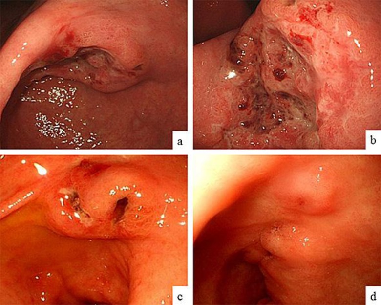
Fig. 2.
Endoscopic findings before chemotherapy and 25 months after chemotherapy. EGD before chemotherapy revealed a Bormann type 3 advanced tumor about 30 mm in diameter in the lower part of the stomach (a). Closer view of the gastric tumor (b). EGD performed 8 months after chemotherapy showed a remarkable reduction in the size of the tumor, which had the appearance of an excavated lesion with marginal protrusion (c). EGD performed 25 months after chemotherapy revealed an even greater reduction in the size of the tumor, which had the appearance of an extremely small elevated lesion with a scar (d).

