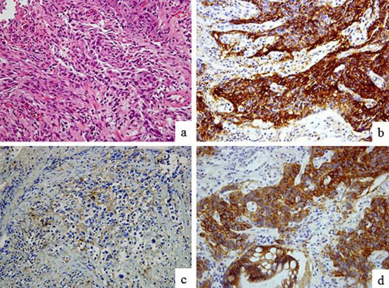
Fig. 3.
Hematoxylin and eosin (HE) staining and immunohistochemical findings of the gastric tumor biopsy specimen. HE staining revealed that the tumor was a poorly differentiated adenocarcinoma (a). Immunohistochemical evaluation of a tumor biopsy specimen revealed that the tumor cells were positive for AFP (b), PIVKA-II (c), and HER2 (d). Original magnification ×400 (a–d).

