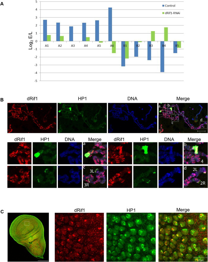
Figure 7. dRif1 regulates timing of replication initiation from origins and associates with HP1.
A) S2 cells untreated (blue bar) or dRif1 RNAi treated (green bar) cells were arrested with HU and labeled with BrdU immediately for 1 hr early replication samples (E) or after 4 hours for late replicating samples (L). The y-axis represents log2E/L value, where log2 of the ratio of enrichment of early vs late for each locus is plotted. X-axis shows the selected euchromatic origins (A1-6) and heterochromatic origins (B1-5) from chromosome 2 and 3. A1-A2 and B1 –B3 are from 2L, A3-A4 and B4 are from 3L, A5-A6 and B5 are from 3R chromosomes. B) Polytene chromosome from the 3rd instar larvae stained for dRif1 (red) and HP1 (green) showing dRif1 localization on multiple regions on polytene chromosomes. Magnified insets (–a-d) are heterochromatic regions (chromo-center and telomere) with intense HP1 foci and dRif1 co-localizes with HP1 in these regions. Arrow heads in the red panel shows that dRif1 localizes to the polytene telomere cytologically. The scale bar represents 20 μm in the polytene spread and 5 μm in the magnified insets. C) dRif1 (red) co-localizes with HP1 (green) in larval wing imaginal disc. The scale bar represents 100 μm in the lower magnification image and 10 μm in the higher magnification image.

