Table 1.
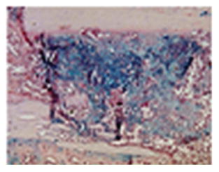
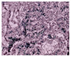
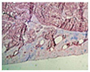
Stages of fracture healing in areas of endochondral bone formation as related to the different time points investigated by FT-IRI in the mouse. Intramembranous bone formation directly results in woven bone and hard callus without prior cartilage formation (stage 1 is excluded since this stage the fracture callus is an amorphous hematoma).
| Stage 1 inflammation | Stage 2 soft callus | Stage 3 hard callus | Stage 4 remodeling | |
|---|---|---|---|---|

|

|

|
||
| Time | Before week 1 | Weeks 1 and 2 | Week 2 and 4 | Weeks 4 and 8 |
| Predominant cell type | Inflammatory cells, platelets, macrophages | Chondrocytes, fibroblast, mesenchymal progenitors | Osteoblasts, “chondroclasts” | Osteoclast, osteoblast |
| Matrix | Hematoma, granulation tissue | ECM proteins (collagen II, Collagen X) | mineralized bone matrix, collagen I, woven bone | Lamellar bone, cortical and trabecular structure |
| Fracture healing processes | Reorganization migration of MSCs | Endochondral ossification, matrix mineralization | Vascular invasion, replacement of cartilage by bone | Bone and matrix degradation, new bone formation |
