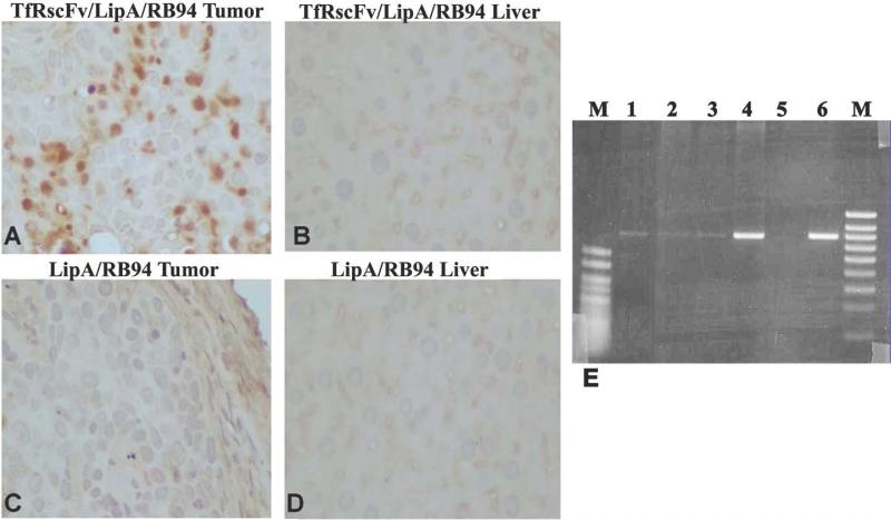
Figure 4. In vivo expression of RB94 in tumor and liver at the single cell level by immunohistocemical analysis and PCR.
In Panels A-D, HTB-9 tumor and liver cells from the mice shown in Figure 3B were immunohistochemically stained for RB94 expression.
Panel A: Tumor from mouse injected with TfRscFv/LipA/RB94 in Figure 3B
Panel B: Liver from the same mouse whose tumor is shown in Panel A.
Panel C: Tumor from a mouse that received the untargeted LipA/RB94 complex in Figure 3B
Panel D: Liver from the same mouse whose tumor is shown in Panel C
Panel E: PCR analysis of tumor bearing bladder, and normal tissues from two individual mice bearing RB94 negative HTB-9 tumors which had been injected three times over 24 hours with the TfRscF/LipA/RB94 complex at 24ugDNA/mouse/injection. Lanes 1-4 are from mouse 1 while lane 6 is from a separate tumor bearing mouse. Lane 1 = liver; Lane 2 = large intestine; Lane 3 = kidney; Lanes 4 and 6 = bladder with tumor; Lane 5 = water blank. M = size markers (500bp and 1000bp hyperladder V and IV, respectively,) (Bioline Co., Randolph, MA).

