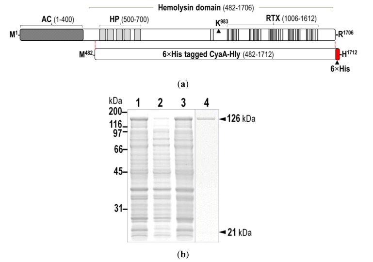
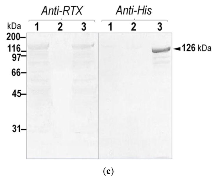
Figure 1.
(a) (Above) Schematic diagram of CyaA showing adenylate cyclase (AC) and hemolysin (Hly) domains. Five putative helices, α1–α5, in the hydrophobic region (HP, residues 500–700) are represented by gray blocks. The palmitoylation site is indicated by Lys983, whereas the repeat in toxin (RTX) region (residues 1006–1612) is represented by multiple lines, with each line corresponding to a single nonapeptide repeat (X-U-X-Gly-Gly-X-Gly-X-Asp). (Below) Diagram of 6×His-tagged CyaA-Hly showing residues 482–1706 with the joining 6×His residues of 1707–1712; (b) SDS-PAGE (12% gel) stained with Coomassie Brilliant Blue of lysates extracted from E. coli (~106 cells) harboring: lane 1, pCyaAC-PF with IPTG induction; lanes 2 and 3, pCyaAC-PF/H6 without and with IPTG induction, respectively. Lane 4 represents the Ni-NTA purified 6×His-tagged CyaA-Hly toxin. Protein bands of CyaA-Hly (~126 kDa) and its activator CyaC-acyltransferase (~21-kDa) are arrowed; (c) Western blot analyses of the corresponding gels from (b) incubated with anti-RTX (left panel) or anti-6×His epitope tag (right panel) antibodies, showing the reacted bands of 126-kDa CyaA-Hly as arrowed.


