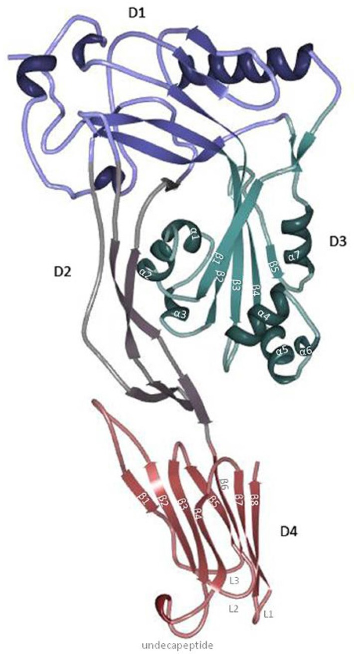
Figure 1.
Perfringolysin O’s (PFO) structure. PFO is dominated by β-strands and is divided into four domains. Domain 4 (red; D4) consists of two β-sheets of four β-strands (D4 β1–4 and D4 β5–8) packed together in a β-sandwich structure connected by four loops (L1, L2, L3 and undecapeptide). Domain 3 (green; D3) contains one core β-sheet (D3 β1–5) flanked by two sets of three α-helices (D3 α1–3 and D3 α4–6) and an additional α-helix (α7) that connects β5 with domain 1 (blue; D1). Domain 1 and domain 2 (purple; D2) connect D3 and D4. D2 is elongated and contains a β-sheet. D1 consists of a β-sheet and four α-helices. The figure was made with RCSB PDB Protein Workshop 4.1.0 (RCSB Protein Data Bank, Piscataway, NJ and La Jolla, CA, USA, 2014) and adapted with Adobe Photoshop CS3 extended (Adobe Systems Incorporated, San Jose, CA, USA, 2007).

