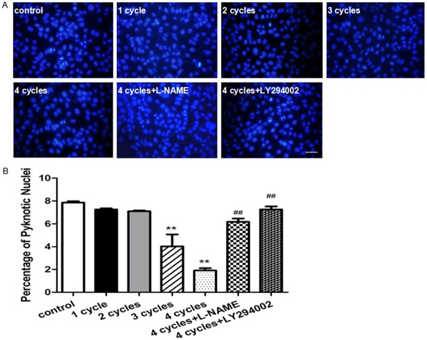
Figure 6.
PEMF protects the apoptosis of HUVECs in vivo. Representative images of Hoechst33258 Staining in HUVECs were stimulated by PEMF for 1-4 cycles, and 4 cycles group treated with L-NAME or LY294002 and stained by Hoechst33258 (A) Quantitative analysis was represented as the apoptotic cells in the total cells per field (B) Values are mean ± SEM; n = 4, N.S. means no significant difference, ** means P < 0.01, vs. control; ## means P < 0.01, vs. 4 cycles group. Scale bar indicated 20 μm.

