Figure 1.

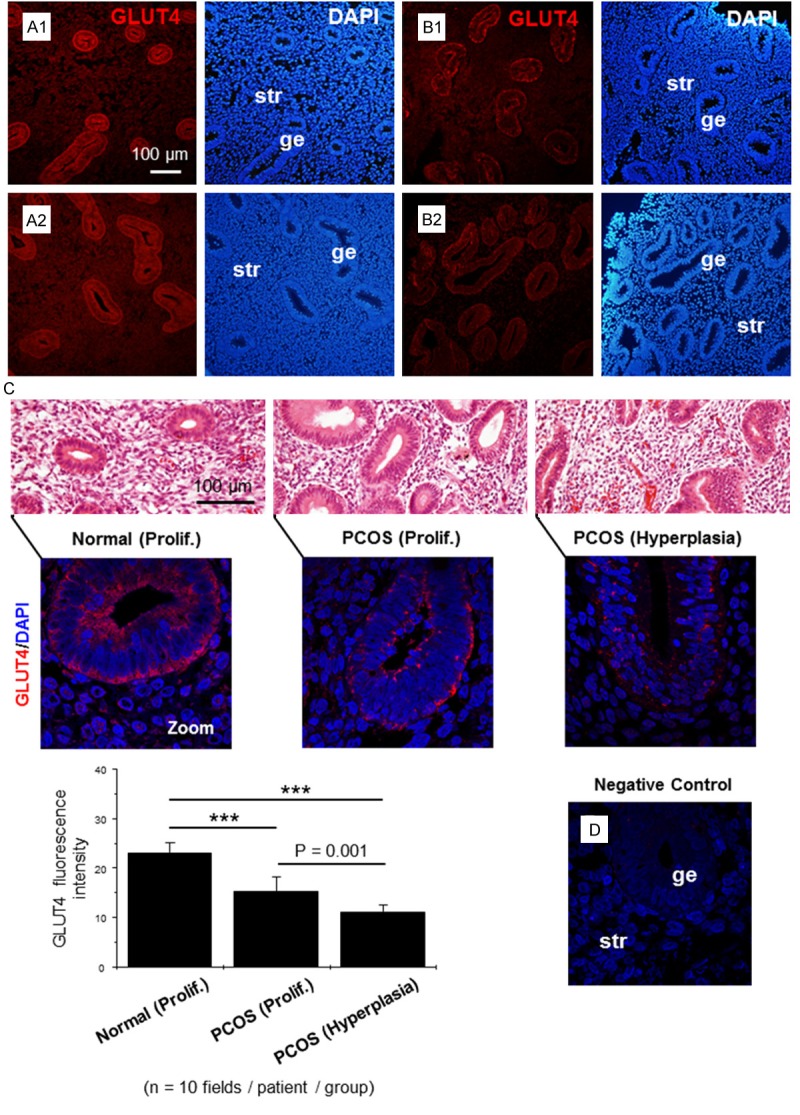
Immunofluorescence localization of GLUT4 in human endometrium. Representative paraffin-embedded endometrial sections in the proliferative stage of healthy women (A1, A2) and women with PCOS (B1) and in women with PCOS and hyperplasia (B2). GLUT4 was significantly decreased in glandular epithelial cells in women with PCOS (B1) and PCOS with hyperplasia (B2) compared to controls (A1, A2). The images are representative of those observed in numerous sections from multiple endometrial tissues. (C) Quantification of GLUT4 immunofluorescence intensity in human endometrium. The top row of images shows the histology of hematoxylin/eosin-stained human endometrial biopsy samples. The lower row consists of magnified images of the top row and shows immunostaining of GLUT4 (red) mainly in the membrane and cytoplasm. Ten fields were observed per patient in each group. Values are the mean ± SD, and significance was tested by one-way ANOVA with Bonferroni correction for multiple comparisons when appropriate. Experiments were performed using different endometrial donors with similar results. The image in the lower right shows the negative control. ***p < 0.001. Prolif., the proliferative phase; ge, glandular epithelial cells; str, stromal cells.
