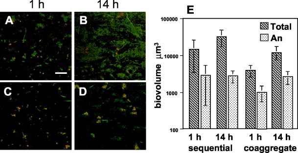
FIG. 5.
The presence of A. naeslundii in four-species biofilm using FISH with Cy3-labled probes. (A to D) Confocal micrographs of mixed-species biofilms hybridized with an A. naeslundii-specific probe (red) and counterstained with general nucleic acid stain Syto 9 (green). Colocalization of both probes appears yellow. (A) One-hour sequentially inoculated biofilm. Bar, 40 μm. (B) Fourteen-hour sequentially inoculated biofilm. (C) One-hour coaggregate-inoculated biofilm. (D) Fourteen-hour coaggregate-inoculated biofilm. (E) Graph indicating the total biovolumes of the four-species biofilms (hatched bars) and biovolumes of A. naeslundii (An) (stippled bars) in sequentially and coaggregate-inoculated biofilms.

