Figure 4.

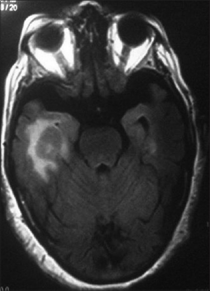
Magnetic resonance imaging showing a homogenously enhancing solid mass lesion abutting right temporal horn of lateral ventricle and a small enhancing lesion in the left temporal lobe

Magnetic resonance imaging showing a homogenously enhancing solid mass lesion abutting right temporal horn of lateral ventricle and a small enhancing lesion in the left temporal lobe