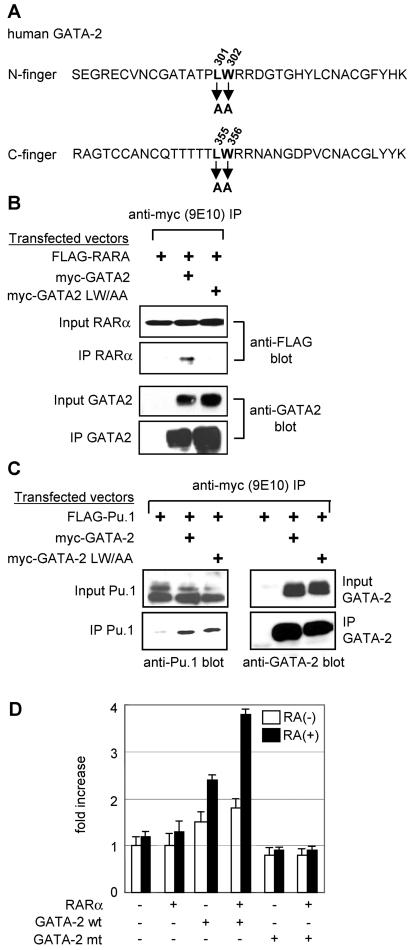
FIG. 7.
Analysis of a GATA-2 mutant that does not interact with RARα. (A) Amino acid sequences of GATA-2 N and C finger regions showing the positions of the mutagenized LW residues. (B) Coimmunoprecipitation analysis of mutant GATA-2 and RARα. 293T cells were transfected with the expression plasmids indicated. Cell lysates were prepared 24 h later, immunoprecipitated (IP) with anti-FLAG antibody, and analyzed by Western blotting with anti-FLAG and anti-GATA-2 antibodies. Input nuclear extracts were analyzed as a control for the levels of proteins expressed. (C) Coimmunoprecipitation analysis of mutant GATA-2 and Pu.1. Experiments were carried out essentially as described for panel B. (D) RA responsiveness of mutant (mt) GATA-2 activity. 293T cells were transfected with a GATA-dependent luciferase reporter plasmid, as well as the indicated combinations of expression vectors for RARα and mutant and wild-type (wt) GATA-2. Cells were then treated with RA (1 μM; solid bar) or diluent alone (open bar) 24 h after transfection, and the luciferase activities were measured another 24 h later. Luciferase activities are standardized against Renilla luciferase activity from a cotransfected control reporter (pRL-CMV-Renilla luciferase) and expressed as fold increases over the activity of the reporter alone. The data presented represent two independent experiments, each of which was performed in triplicate.

