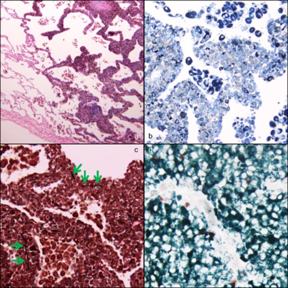
Figure 3.

(a) Hematoxylin–eosin staining (×40) showing atypical cells with melanin granules proliferating in a lepidic-like fashion, simulating lepidic predominant adenocarcinoma (in the right half). (b) S-100 staining (×200) shows that the atypical cells are positive (appear gray). (c) Elastic van Gieson staining (×200) shows that the elastic membranes of the alveolar septa are intermittently disrupted (green arrows). (d) Ki-67 staining with May-Giemsa staining (×400) shows that brown and dark green nuclei are positive, but white ones are negative. The Ki-67 labeling index, average proportion of positive cells in three hot spots of the tumor, was 33.4%.
