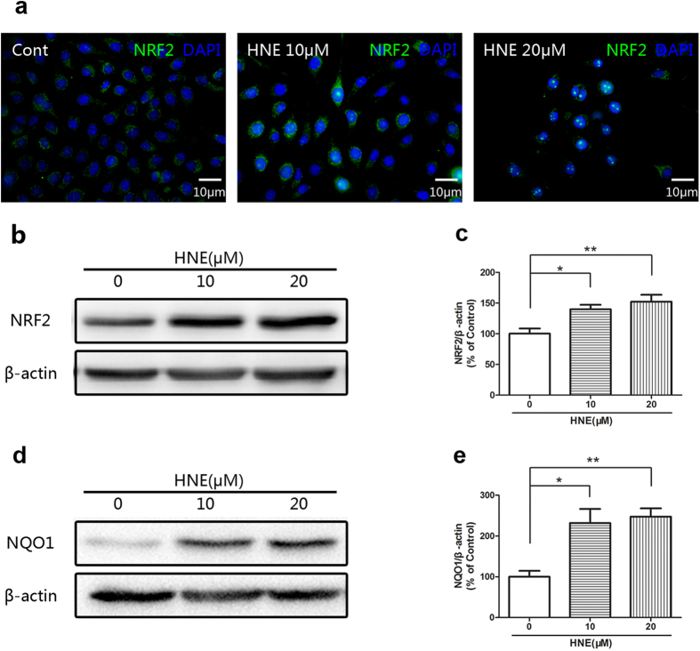
Figure 4. 4-Hydroxynonenal induced the activation of NRF2 in the cultured human corneal epithelial cells (HCE).
a: Representative images of immunofluorescent staining with anti-NRF2 antibody after treatment of 4-Hydroxynonenal (10 and 20 μM) for 24 hours. Green color: NRF2; Blue color: nuclear DAPI staining. Scale bar: 10 μm. b: Representative images of Western blot with anti-NRF2 antibody after treatment of 4-Hydroxynonenal (10 and 20 μM) for 24 hours. c: Statistical analysis of Western blot results of NRF2. Data were presented as mean ± SEM; n = 10; *: p < 0.05, **: p < 0.01. d: Representative images of Western blot with anti-NQO1 antibody after treatment of 4-Hydroxynonenal (10 and 20 μM) for 24 hours. e: Statistical analysis of Western blot results of NQO1. Data were presented as mean ± SEM; n = 4; *: p < 0.05, **: p < 0.01. (HNE=4-Hydroxynonenal).

