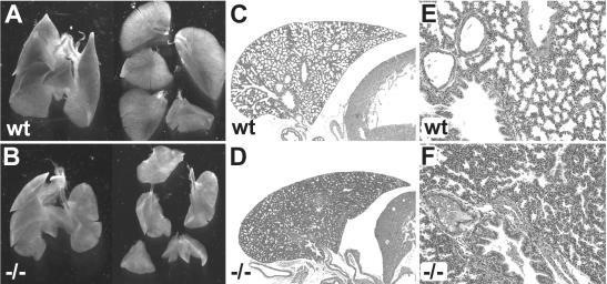
FIG. 4.
Lung hypoplasia in Sox11-deficient mice. (A and B) Appearance of the lung and the five separate lung lobes at 18.5 dpc. In counterclockwise orientation starting at the top, the lobes are as follows: cranial right lobe, middle right lobe, caudal right lobe, accessory right lobe, and left lobe. (C to F) Hematoxylin-eosin staining of paraffin-embedded sections of the left lobe at low (C and D) and high (E and F) magnifications. (A, C, and E) Wild-type embryo; (B, D, and F) severely affected Sox11-deficient littermate. Note that neither embryo had breathed and that the lungs had not been inflated with air.

