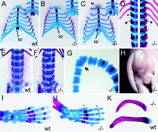
FIG. 8.
Skeletal malformations in Sox11-deficient embryos. (A to C) Alizarin red (bone) and alcian blue (cartilage) staining of rib cages (ventral view) of wild-type (wt) embryo (A) and Sox11-deficient (−/−) littermates (B and C) at 18.5 dpc. Insets show magnifications of the caudal sternum. xp, xiphoid process of sternum. (D) Alizarin red (bone) and alcian blue (cartilage) staining of spinal column (dorsal view) of Sox11-deficient embryos at 18.5 dpc at thoracolumbar level. The asterisk marks the missing 13th rib. Arrows point to additional rib studs on the first lumbar vertebra. Two spinal columns are shown, with each being half-sided. (E and F) Lumbar spinal columns of wild-type (wt) and Sox11-deficient embryos at 18.5 dpc from the ventral aspect showing malformations of vertebral bodies. (G and H) Kinked tail of Sox11-deficient mouse at 18.5 dpc shown by alizarin red and alcian blue staining (G) and by outer appearance (H). The arrow in panel G points to the fused caudal vertebrae. (I and J) Hindlimbs of wild-type (wt) and Sox11-deficient (−/−) mice at 18.5 dpc showing reduced bone formation of talus (white arrow in panel I) and phalanges (black arrowheads in panel J). (K) Clavicles from Sox11-deficient (−/−) (top) and wild-type (wt) (bottom) siblings at 18.5 dpc showing abnormal ventral curvature of the Sox11-deficient clavicle.

