Figure 4.

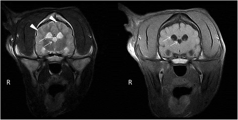
Transversal MRI T2W, T1W images. A symmetrical, bilateral ventriculomegaly of the lateral and the third ventricles (arrows), with laminar hyperintensity visible on T2W images on the border of the grey and white matter (arrowheads).

Transversal MRI T2W, T1W images. A symmetrical, bilateral ventriculomegaly of the lateral and the third ventricles (arrows), with laminar hyperintensity visible on T2W images on the border of the grey and white matter (arrowheads).